
Ultrasound and Echo in Hialeah
Ultrasound and echocardiography use sound waves to create images of the heart, organs, and blood vessels without radiation. At Ven A Verte Medical Center, imaging is part of a primary care approach that connects what we see with your symptoms and your overall health.
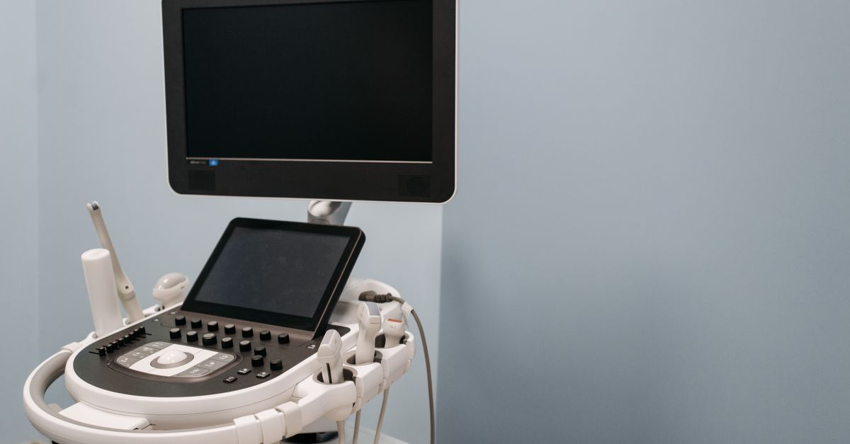
What is Ultrasound and Echo?
Ultrasound, also called sonography, sends high-frequency sound waves into the body and listens for the echoes that return. A computer turns those echoes into real-time images. Because it uses no ionizing radiation, ultrasound is a widely used imaging tool for looking at the abdomen, pelvis, thyroid, blood vessels, and soft tissues.
An echocardiogram is an ultrasound of the heart. It shows the size of the heart chambers, how well the muscle pumps, how the valves open and close, and whether there is fluid around the heart. Echo is commonly used to evaluate shortness of breath, swelling, murmurs, and to follow known heart conditions over time.
These studies help adults of many ages, including people with high blood pressure, diabetes, or a family history of heart disease. Medical guidelines recognize ultrasound as a safe, noninvasive way to answer many clinical questions quickly, often guiding the next step in care without the need for more invasive testing.
Common reasons for imaging
- Chest discomfort, shortness of breath, or unexplained fatigue
- Swelling in the legs, ankles, or feet
- Heart murmur or irregular heartbeat found during an exam
- Abdominal pain, bloating, or concerns about the liver, gallbladder, or kidneys
- Thyroid nodules or a lump in the neck
- Pelvic pain or other concerns involving pelvic organs
- Leg pain or swelling that raises concern for a blood clot
What to Expect
- Uses sound waves rather than ionizing radiation
- The scan itself is typically painless and often takes under an hour
- Preparation instructions are given when the study is ordered
- Results are reviewed and explained by your clinician
- Follow-up imaging or referral is discussed when appropriate
Our Approach to Ultrasound and Echo
Initial Assessment
We begin with your history, symptoms, and a physical exam. This helps decide whether imaging is useful and which type of study best answers your question.
Personalized Plan
Your clinician explains the study, gives preparation instructions, and ties the images to labs and other findings so the plan fits you.
Ongoing Support
Findings are reviewed with you, and follow-up imaging or referral to a specialist is discussed when it is appropriate.
Benefits
No Radiation
Ultrasound uses sound waves rather than ionizing radiation, which is why it is widely used for many types of evaluation.
Safe and Noninvasive
Medical guidelines recognize ultrasound as a safe, noninvasive way to answer many clinical questions quickly, often guiding the next step in care without the need for more invasive testing.
Connected to Your Care
Your clinician ties the images to labs and other findings so the plan fits you.
What to Expect
First Visit
Discuss your symptoms and history. If imaging is appropriate, you receive preparation instructions. The scan itself is typically painless and often takes under an hour.
Week 2-4
Results are reviewed and explained. Some findings are reassuring; others call for added testing or a treatment plan.
Month 1-3
Any recommended changes are put in place, and follow-up is arranged to see how you are responding.
Long-term
If you have a condition that needs monitoring, repeat imaging may be used at intervals your clinician recommends.
Types of studies and how they are used
Depending on your needs, imaging may include a transthoracic echocardiogram of the heart, abdominal ultrasound for the liver, gallbladder, kidneys, and spleen, thyroid ultrasound, pelvic ultrasound, or vascular ultrasound to look at blood flow in the arteries and veins.
Imaging is one tool among many. Results may lead to lifestyle changes, medication adjustments, additional testing, or a referral, always decided together with you. Ask our Hialeah team which studies are available at the office.
Common Questions About Ultrasound and Echo
Is an ultrasound painful?
Ultrasound is generally comfortable. A technician moves a handheld probe over gel on the skin. You may feel light pressure, and some areas of the abdomen can feel tender if they are already sore.
What is the difference between an ultrasound and an echo?
An echocardiogram, or echo, is an ultrasound focused on the heart. It shows the heart chambers, valves, and pumping function. Other ultrasounds look at organs, vessels, the thyroid, or soft tissue.
Do I need to prepare?
Preparation depends on the exam. Abdominal studies may require fasting for several hours, and pelvic studies may need a full bladder. Our team will give you instructions specific to your study when it is ordered.
Does ultrasound use radiation?
No. Ultrasound uses sound waves rather than ionizing radiation, which is why it is widely used for many types of evaluation.
How will I get my results?
Images are reviewed and your clinician explains what they show and what the next steps are. Call (786) 542-1882 to ask how results are shared at our Hialeah office.
Ready to Get Help with Ultrasound and Echo?
Call (786) 542-1882 or visit us at 5341 W 20th Ave, Hialeah, FL 33012.
Book an Appointment